Lab activity : Extreme necessity of vascular tissues
I- Vascular tissues of the celery
Goals:
- 1. To observe the structure of vascular tissue in plant stems, roots, and leaves.
- 2. To determine the function of phloem and xylem tubes in vascular plants.
Background Information:
Vascular plants first appear in the fossil record about 410 million years ago.
Vascular plants are plants with tube-like cells that make up vascular tissue. The tubes are constructed of lines of interconnecting cells. These tubes serve as a means of transport for food, minerals, water, and other materials within the more advanced plants.
Of course, this method of transport is a much more sophisticated system than the cell-to-cell system used by more primitive plants like mosses and liverworts. (These more primitive plants are called non-vascular.)
Because of the development of vascular tissue, vascular plants can grow very large.
Phloem and xylem cells are the two main components of vascular tissue. (Other components are fiber cells for strength and storage cells for storage.) Phloem tubes transport food that is made by the leaf during photosynthesis. The food is brought from the leaf down to all other parts of the plant. Xylem tubes transport water and minerals from the roots up to the stem and the leaves.
Materials: Prepared slides of vascular tissue: roots, stems, and leaves, food coloring, beaker of water, celery stalks, scalpels, hand lenses,
Procedure:
Complete the following stations. Be sure to answer all questions using complete statements.
STEP 1: Transporting Materials Demonstration
The celery stalks at this station were placed in a vase filled blue coloring.
- Observe the celery stalks and draw what you observed.
- Answer the following questions:
Just to go further :
questions :
- What do you notice about the leaves at the top of the celery stalks?
- How the dye made its way to the top of the stem?
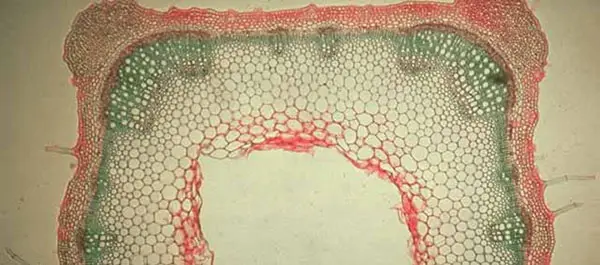
STEP 2: Vascular Tissue Slide under the light microscope
Look at the prepared slide of a stem cross-section under the microscope. The stain used here is iodine green carmine. This technique reveals green lignified tissue and pink non-lignified tissue.
Choose the most appropriate magnification for the best view and take a photo to add to your answers.
question :
- What is vascular tissue and what is it used for?
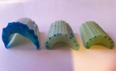
STEP 3: Vascular Bundles
Lateral View (Horizontal View)
- Cut off a small, lateral/horizontal cross section of the celery stem.
- Using a hand lens observe the small vascular “bundles.” They were stained blue by the dye.
- Take a photo and label a vascular bundle.
Complete the following sentences:
1. What I Observed:
2. What I Learned:
3. What I Wonder:
II- the motor of xylem
To analyse on the next Week
According to the transpiration-adhesion-cohesion-tension mechanism of water transport in xylem, water evaporating through the stomata produces a tension, or negative pressure, that pulls the water column up the plant. This column is maintained by the hydrogen bonding between water molecules (cohesion) and the hydrogen bonding between water molecules and the molecules that make up the cell walls (adhesion).
If the above mechanism is correct, then a twig that has been removed from the rest of the plant and attached to an artificial water column (as in a pipette) could pull water up the pipette. The rate of movement can be determined by adding a dye to the water at the bottom of the pipette (like methylene blue).
Materials:
Begin Experiment: each group need 2 cherry laurel twigs.
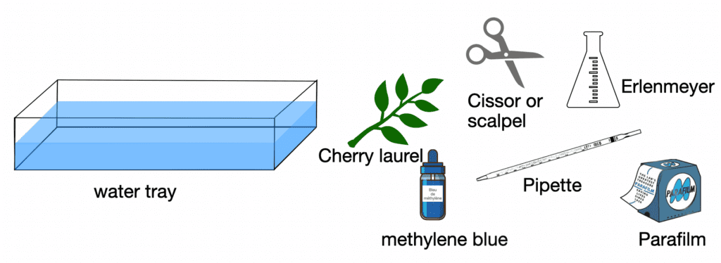
Procedure:
A- Place a pipette under water using the large water tray. Manipulate the pipette until it is completely filled with water – WITHOUT air bubbles.
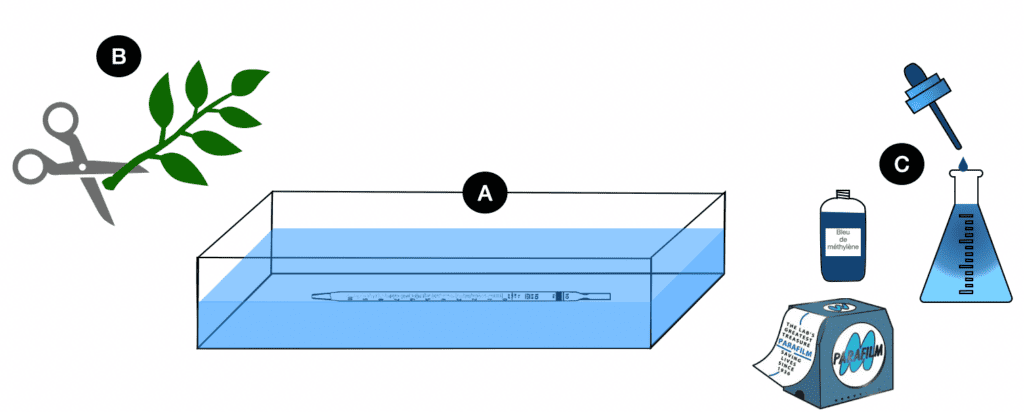
B- Place the cut end of the cherry laurel twig under water and cut off another centimeter to insure that there are no bubbles in the transport tissue of the plant that would disrupt water transport.
C- Fill the Erlenmeyer flask with concentrated methylene blue and homogenise the dye.
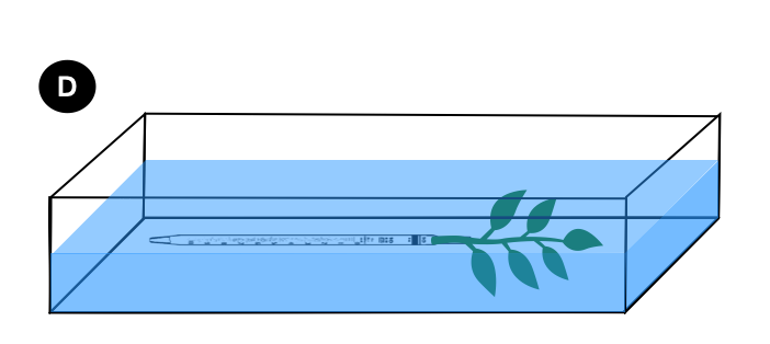
D- Then place the cherry laurel twig into the water tray. Cut off an additional cm of stem under water. Then insert the twig into the tubing. For our purposes, it has to be fitted snugly. Insert the freshly cut end into the above tubing while under water, making certain that NO air bubbles are trapped within the assembly. If the twig does not fit tightly in the pipette, wrap several strips of Parafilm around the stem before inserting it into the tubing.
E- For your stalk with leaves : by placing your finger on the tip of the pipette, transfer the whole so that the tip is now in the dye.
Transfer everything to a stand to hold the device and position it in the dye, without losing any water. Don’t forget to place everything in the light
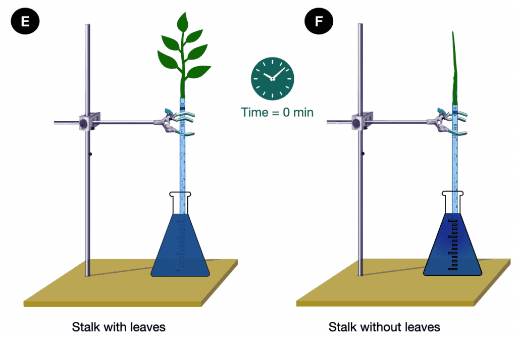
F- This same set-up is repeated with a cherry laurel stem without leaves to use as a control.
=> Thus, next week, you can see whether or not the dye has risen.
Vocabulary :
Pour en savoir plus: University of Georgia – different tissues