Lesson: Basics in biology : DNA

Many people believe that James Watson and Francis Crick discovered DNA in the 1950s. But, this is not the truth. Firstly, DNA was identified in the late 1860s by a chemist Friedrich Miescher. Then, the race was on to discover the molecular structure and its coding by other scientists (notably, by Erwin Chargaff). These efforts led to the convergence of details of the molecule, enabling Watson and Crick to discover the double helix and its detailed structure in 1953.
I- The organisation of living organisms
A- The scales of life
All multicellular organisms are individuals within their species. It is made up of organs (e.g. heart, liver, lungs, etc.) often representing functional systems or parts (circulatory system, digestive system, respiratory system, etc.) that perform the major functions of the organism.
Doc 1: scales of cell organisation
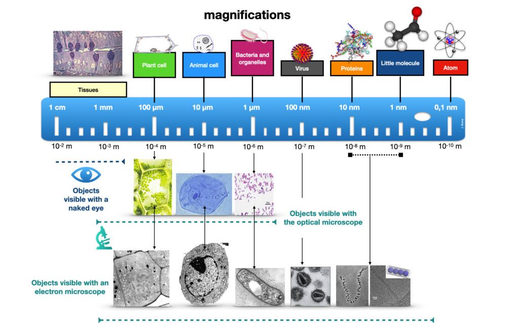
To test a scale : https://learn.genetics.utah.edu/content/cells/scale/
Each organ is made up of tissues representing the different parts of the organ and performing specific functions. Each tissue will then have cells specialised in these same functions and will therefore have a variable organelle content, depending on its specialisation. Finally, each cell will have a particular wealth of molecules that will differ from another cell with a different speciality.
The cells of multicellular organisms are generally joined together and surrounded by an extracellular matrix that enables cells to adhere and tissues to bind together. For example, plant cells are surrounded by a cell wall made up of various molecules including cellulose and pectins.

Definition: Extracellular matrix: The extracellular matrix is a set of large molecules present in tissues but located outside the cells that synthesise and secrete them. The extracellular matrix facilitates bonding and adhesion between cells and organises them into tissues.
B- Tools for observing living organisms
With an optical microscope, we can observe the tissues that make up organs and the cells that make up tissues, but the maximum magnification is around x1000.
With an electron microscope, we can observe more details, including organelles. There are 2 techniques: one that allows us to see the inside of the cell, transmission electron microscopy (TEM), and the other that allows us to see the cells in relief, scanning electron microscopy (SEM).
Like mineral matter, living matter is made up of molecules, assemblies of atoms that are too small to be observed using microscopy (nm).
Be aware: Don’t confuse cells and molecules!

A cell measures between 100 and 2 µm. But a molecule is much smaller. It is an assembly of atoms. A cell is made up of a multitude of molecules, and a molecule of different atoms. But confusing a molecule with a cell is a bit like confusing a city with a country.t surrounded by a cell wall made up of various molecules including cellulose and pectins.
II- The importance of the information carried by DNA
DNA or Deoxyribonucleic Acid is the molecule that carries genetic information. It is a molecule that is contained in the nucleus (in the form of chromatin) when it is in its readable form. When the cell is dividing, however, the nuclear envelope disappears and the DNA condenses to take the form of chromosomes.
But in a specialised tissue cell, not all the genes are expressed, and only a part corresponding to the specialisation is expressed.
A- The structure of the DNA molecule
Reminder: DNA = DeoxyriboNucleic Acid.
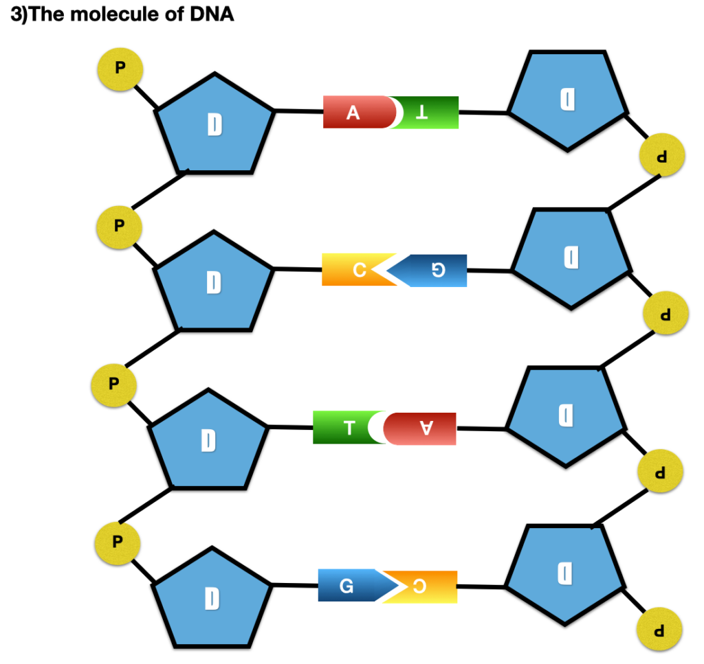
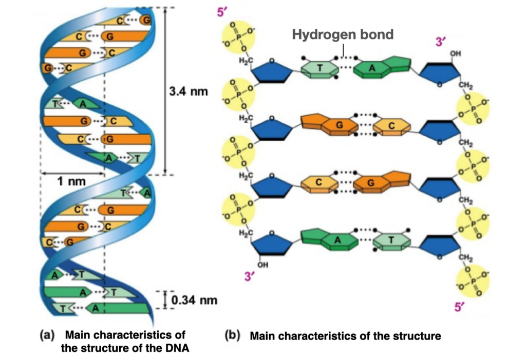
This molecule is made up of two chains wound around each other in a double helix (for this reason it is called double stranded = 2 chains). It is a polymer made up of a succession of elementary molecules called nucleotides. There are 4 possible nucleotides in DNA:
Doc 2: scales of cell organisation
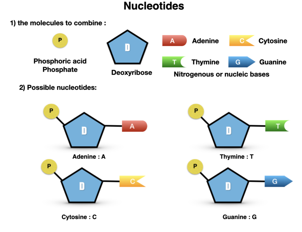
These nucleotides link together to form a long helix-like chain. The second chain is complementary to the first according to the following rule: A with T and G with C.
The structure of the DNA molecule is universal in the living world.
B- The language of DNA
A succession of nucleotides is called a sequence, because it forms a succession of letters. If this succession of letters results in a coherent sentence, which enables the synthesis of a protein, it is called a gene… Similarly, if a gene enables the production of a particular protein with a specific function, there may be different versions of the same gene, resulting in specific or non-specific modifications to the protein. These different versions of the same gene are called alleles.
Doc.3: Example of the insulin gene sequence:
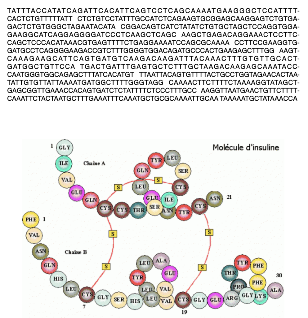
The nucleus of all cells contains absolutely all the DNA that characterises a multicellular organism, but not all the genes are expressed. So, depending on the specialisation of the cell, only certain genes useful for specialisation are expressed. In conclusion, depending on the cell’s specialisation, some genes are active and others are rendered silent.

Definition: A gene is a segment of DNA which contains the information required to synthesise a protein. The gene is said to be active/on/expressed when this synthesis takes place. Otherwise, it is inactive/repressed. But obviously, gene expression is not a black and white process: there are plenty of grey levels, with, for example, genes that are very active, over-expressed (high synthesis) or even partially repressed (very low synthesis)…
Conclusion:
All the cells of a multicellular organism are the result of successive divisions of the egg cell at the origin of the organism. They all contain the entire genome, but express only part of it. So, throughout life, cells use certain genes to produce their own molecules according to their speciality: genes are said to be expressed. By specialising, cells acquire differences that distinguish them (more than 250 cell types have been identified in the human species).
To go further: ask a biologist


